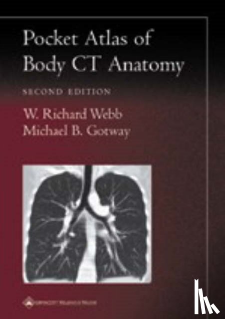
Featuring 229 images, this atlas is a guide to interpreting computed tomography body images. It shows readers to recognize normal anatomic structures on CT scans and distinguish these structures from artifacts. Chapters in this book cover the neck and larynx, thorax, portal venous phase abdomen, pelvis, arterial phase abdomen, and reconstructions.
I have a question about the book:
'Pocket Atlas of Body CT Anatomy - Webb, W. Richard, Gotway, Michael B.'.
Fill in the form below.
We will respond as fast as possible.
We value your privacy
We use cookies to measure traffic, improve Boekstra and let Google tailor ads to your interests. You can keep using the site either way — even if you decline. More info